
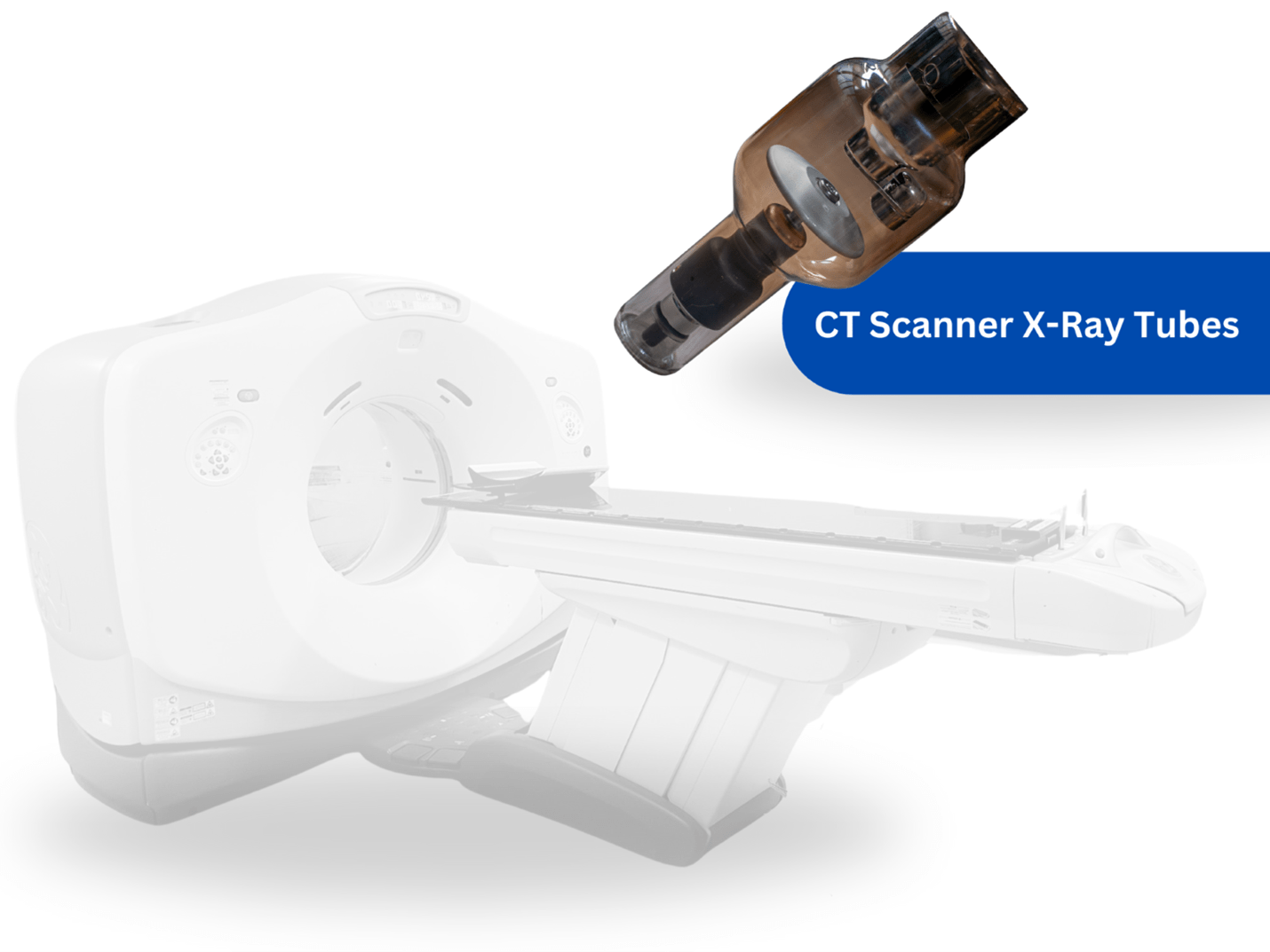
Introduction When it comes to maintaining and replacing CT scanner tubes, healthcare professionals and administrators…

Are You Missing The Added Revenue of Dense Breast Imaging?
Most radiologists are not currently providing a comprehensive dense breast solution. Mammography and other x-ray technologies lack the ability to effectively detect lesions in dense breasts.
What is a Dense Breast?*
Dense breast tissue is comprised of less fat and more connective tissue which appears white on a mammogram. Cancer also appears white thus tumors are often hidden by the dense tissue. As a woman ages, her breasts usually become more fatty. Radiologists have been reporting a woman’s dense breast tissue to her referring doctor for twenty years. Most often, that information is not conveyed to the patient. Displaying heterogeneously or extremely dense breast tissue on a mammogram is considered dense (BIRADS C, D).

Dense Breast Statistics**
- It’s The Law
- Starting April 1, 2013, California law requires that patients be informed if they have “dense breast tissue”
- #1 Health Risk for Women
- Breast cancer is the #1 health risk for women and the second leading cause of death of ALL women in the US
- Approximately 40% of Women Have Dense Breasts
- Approximately 40% of women undergoing screening mammography are classified as having either “heterogeneously dense” or “extremely dense” breasts. For all of these women, the patient letter will inform them that they have “dense breast tissue.”
- Reduce False Negatives***
- In an ASTOUND Study, many cancers are being missed in dense breasts by not using ultrasound in addition to mammography.

Personal Stories
Dr. Nancy Cappello – Was diagnosed with Stage 3c breast cancer six weeks after receiving a “normal”mammography report.
Joan Lunden – Was diagnosed with having Breast Cancer after both a Mammogram and 3D Mammogram showed “normal” test.
Michelle’s Story – Cancer detected at age 44, hidden by dense breasts and would have benefited from an automated ultrasound 3 years earlier.
Brittany’s Story – Age 29, a 1.7 cm tumor detected, hidden by dense breasts. Would have benefited from an automated ultrasound 1 year earlier.
Elaine’s Story – Age 66, a 9.5 cm tumor detected, hidden by dense breasts. Would have benefited from an automated ultrasound 4 years earlier.
If you don’t have a solution for the detection of dense breast cancer, contact us today to find out more about our Robotic Ultrasound Solution. Fill out our online form or email us at info@OncologySystems.com.
RELATED LINKS
ABUS: The Future of Breast Screening
*Are You Dense Organization. 2016. Web.http://www.areyoudense.org
**Breast Density Information. 2016. Web.http://www.breastdensity.info
***Astound Study. Data presented is from the American Society of Clinical Oncology article. 2016. Web. http://jco.ascopubs.org
Related Posts

Comments (0)